
Pancreatic Cancer Is Often Found Too Late. What Can We Learn Earlier?

Advanced imaging, genetic insights, and emerging blood-based tests are creating new opportunities to identify pancreatic cancer risk before symptoms appear. The challenge is knowing what to look for and when to act.

November 18, 2026
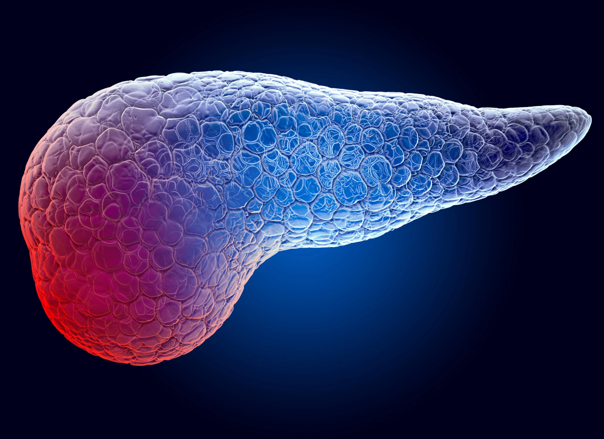
3D illustration of the human pancreas, with the head of the organ highlighted in red.
by Sarah Pesce
Chief Operating Officer, Radence

Pancreatic cancer is one of the most difficult cancers to detect early. Symptoms can be vague or absent until the disease has progressed, and there is currently no broadly recommended screening test for people at average risk.

The consequences are significant. The five-year relative survival rate for pancreatic cancer is less than 15%. When the disease is detected while still confined to the pancreas, that figure rises to approximately 44%.1

These numbers underscore why earlier detection matters. They also raise an important question: What can we learn about pancreatic cancer risk before symptoms appear?

At Radence, our approach to pancreatic cancer early detection begins with a more complete understanding of individual risk. Advanced imaging, whole-genome sequencing, metabolic testing, and family history each provide important information. Our physicians bring these findings together, drawing on continuously evaluated scientific evidence to determine what warrants closer investigation, what can be monitored, and when intervention may be appropriate.

This matters because the significance of a finding is rarely determined by a single test. A small pancreatic cyst, a change in blood sugar, or an inherited genetic variant may take on greater meaning when considered alongside the rest of a person's health information.

Sometimes the First Clue Is Found on an MRI

One of the important advantages of comprehensive imaging is the ability to identify unexpected abnormalities in people who have no symptoms. Radence's BioAtlas whole-body MRI can provide an opportunity to detect incidental findings that might otherwise remain undiscovered.

One example is an intraductal papillary mucinous neoplasm, or IPMN, a type of cyst that develops within the ducts of the pancreas.

IPMNs are increasingly discovered incidentally during MRI and CT scans, often in people who have no symptoms. Most never develop into cancer. However, some have characteristics that make them more concerning, including their size, growth rate, internal structure, and involvement of the main pancreatic duct.

Identifying an IPMN through imaging can bring a previously unknown finding to clinical attention, creating an opportunity for further evaluation before symptoms or complications develop.

There is another reason these findings deserve attention. Research suggests that people with IPMNs can have an increased risk of developing pancreatic cancer elsewhere in the pancreas, even when the original cyst remains stable.

This distinction has important implications for how pancreatic findings are evaluated and monitored over time.

"An IPMN may appear to be a small, isolated finding, but it can tell us something important about risk throughout the pancreas," states Brian Anhalt, Director of Clinical Implementation at Radence. "We look beyond the cyst itself to understand what it means for the individual and how that risk should be monitored over time."

Understanding Risk Requires More Than Imaging

Imaging provides essential information about the pancreas, but it is only one part of a comprehensive evaluation.

At Radence, when an IPMN is identified, our clinical team assesses the finding in the context of the member's broader health profile. This includes several sources of information:

  • Advanced imaging: Our BioAtlas whole-body MRI protocol may identify an incidental pancreatic finding. Dedicated pancreatic MRI, including MRCP, can then provide a more detailed view of the lesion and pancreatic ducts when indicated.
  • Genetic risk: Whole-genome sequencing can identify inherited variants associated with increased pancreatic cancer risk, including certain BRCA-related mutations.
  • Family and medical history: A family history of pancreatic cancer, previous pancreatic conditions, and other established risk factors help inform the evaluation.
  • Metabolic health: Changes in blood sugar regulation, particularly new-onset diabetes after age 50, can sometimes signal an increased risk of pancreatic cancer.

That last connection is especially important.

Research has shown that new-onset diabetes in adults over 50 can precede a pancreatic cancer diagnosis. While the vast majority of people who develop diabetes do not have pancreatic cancer, the change may warrant closer investigation in someone with additional risk factors.2

Radence evaluates metabolic health through testing that includes measures of insulin resistance, which may become abnormal before conventional blood sugar markers such as HbA1c.

By considering these findings together, our physicians can recognize changes that may alter an individual's risk assessment and determine whether additional pancreatic evaluation is appropriate.

Can a Blood Test Help Detect Pancreatic Cancer Earlier?

New blood-based technologies are expanding the possibilities for pancreatic cancer early detection. But incorporating an emerging test into clinical care requires more than promising performance data.

At Radence, our clinical protocols are informed by TechAtlas, the scientific research division of RA Capital, whose physicians and PhD scientists continuously evaluate emerging diagnostics, therapeutics, and biomedical evidence. This scientific foundation helps our physicians assess the strength of the evidence, understand the limitations of new technologies, and determine when they may meaningfully influence care.

One example is Avantect, a pancreas-focused blood test developed by ClearNote Health. It analyzes molecular changes in DNA fragments circulating in the bloodstream that may signal the presence of pancreatic cancer, including earlier-stage disease.

While early research is promising, additional studies are needed to establish its effectiveness in real-world screening and its impact on clinical outcomes.

Radence considers Avantect selectively for members whose risk factors and clinical circumstances make the additional information potentially useful. For example, a member over age 50 who develops new-onset diabetes, particularly alongside an IPMN, inherited susceptibility, or family history of pancreatic cancer, may warrant consideration.

Importantly, Avantect does not replace pancreatic imaging or establish a cancer diagnosis on its own. A positive result may prompt additional imaging or specialist evaluation, while a negative result does not eliminate the need for appropriate surveillance.

"The question we ask about any new diagnostic technology is whether the information will change what we do for a member. A promising test is only useful if we understand its limitations and have a clear plan for acting on the results," explains Brian Anhalt, Director of Clinical Implementation at Radence.

Why Continued Surveillance Matters

An IPMN that remains unchanged for several years may be reassuring. But stability does not necessarily mean that future pancreatic cancer risk has disappeared.

Current clinical guidelines allow surveillance to be discontinued in certain patients with small, stable branch-duct IPMNs after five years. However, long-term studies have demonstrated that pancreatic cancer can still develop beyond that period.

Radence takes a more cautious approach, favoring continued surveillance when clinically appropriate and when a member could benefit from intervention if a concerning change were detected.

The frequency and type of follow-up depend on the individual's risk profile, including imaging findings, genetic predisposition, metabolic changes, and other clinical considerations.

We also work with pancreatic specialists to evaluate findings and determine when additional testing, such as endoscopic ultrasound or molecular analysis of cyst fluid, may help clarify risk.

The objective is to detect meaningful changes while avoiding unnecessary procedures. Most pancreatic cysts will never become cancerous, and more aggressive testing can carry real consequences, including false-positive results, anxiety, and potentially avoidable interventions.

Knowing when to investigate further is as important as knowing when continued observation is the appropriate course.

Earlier Insight. Informed Decisions. Continued Attention.

The science of pancreatic cancer early detection continues to evolve, creating new opportunities to recognize risk and investigate concerning changes before symptoms develop.

At Radence, our clinical team draws on advanced diagnostics, rigorous scientific evaluation, and collaboration with leading specialists to understand what those findings mean for each member. As new information emerges, we revisit risk, refine surveillance plans, and determine when further action is warranted.

A pancreatic cyst discovered today may remain stable for years. Or changes in imaging, metabolic health, or other clinical findings may call for a closer look. What matters is having a physician-led team that understands the full picture and stays engaged as it evolves.

Because finding something early only creates an opportunity. Knowing what to do next is what makes that opportunity meaningful.

References

[1] National Cancer Institute. Cancer Stat Facts: Pancreatic Cancer. Surveillance, Epidemiology, and End Results (SEER) Program. Survival statistics based on 2016–2022 data.

[2] Sharma A, Kandlakunta H, Nagpal SJS, et al. Model to Determine Risk of Pancreatic Cancer in Patients With New-Onset Diabetes. Gastroenterology. 2018;155(3):730–739.e3.

A More Complete Understanding of Your Health

Radence brings together advanced diagnostics, scientific expertise, and ongoing physician-led care to help you understand your health risks and make informed decisions about what comes next.
Discover Radence Membership

The Radence Standard

A different standard than the market is applying - and it will always be the one we hold. If you've been considering membership, we'd welcome the conversation.
Sarah Pesce - Radence COO
Sarah Pesce
Chief Operating Officer, Radence
Sarah Pesce, ANP, MSCDL, is the Chief Operating Officer at Radence. She previously served as COO of Thorne Lab, a division of Thorne HealthTech, where she led the development of a clinical platform offering a comprehensive, 360-degree view of individual health. Prior to Thorne, Sarah was COO of Lab100 at Mount Sinai Medical Center, where she shaped clinical operational strategies for advanced digital screening assessments. She also served as Senior Director of Business Development and Clinical Operations for Mount Sinai’s Executive Health Program and worked as Nurse Practitioner for Dr. Valentin Fuster, managing complex cardiology patients and launching impactful ambulatory programs.

You may be interested in:

Not All Whole-Body MRI Scans Are the Same

At Radence, advanced imaging is a critical part of how we look for disease before symptoms appear. That requires more than access to an advanced MRI scanner. It requires a protocol designed to capture the level of detail needed to identify subtle findings consistently.

That is why we developed BioAtlas, our proprietary whole-body MRI protocol. Every aspect is intentional, from how images are captured and which views are included to who interprets them and how the findings are evaluated alongside other health data.

The result is a more detailed and clinically useful view of your health, designed to give your clinical team better information to determine what deserves attention and what should happen next.

BioAtlas also sits within a broader Radence imaging strategy. When MRI cannot fully answer a clinical question, we incorporate other imaging modalities based on your biology, risk factors, and findings.

The result is a more detailed and clinically useful view of your health, designed to give your clinical team better information to determine what deserves attention and what should happen next.

A Whole-Body MRI Is Only as Good as the Protocol Behind It

Two whole-body MRI scans can be performed on similar scanners, take a similar amount of time, and look nearly identical to the person being scanned, yet provide very different information to the clinical team.

MRI protocols contain hundreds of technical decisions that influence what can be seen and measured: the sequences captured, image resolution, anatomical coverage, slice thickness, coil selection, timing, and even how you are positioned within the scanner. Small differences can affect image quality or determine whether specific quantitative information can be extracted from the exam.

Think of it like photography. Two photographers can use the same high-end camera and photograph the same subject, yet differences in the lens, focus, lighting, exposure, and composition can produce very different results.

That is why Radence defines BioAtlas at a detailed technical level and works directly with our imaging partners to standardize how the protocol is installed, tested, and performed.

What the Scan Doesn’t Capture, You Can’t Interpret Later

Think of an MRI as creating a series of slices through your body, much like slices in a loaf of bread. If there are gaps between the slices, portions of the anatomy are never imaged. If the slices are too thick, subtle abnormalities can become blurred or partially hidden.

BioAtlas was developed with input from leading national experts, across academia institutions and community leaders, with decisions around slice thickness and coverage made intentionally to balance fine anatomical detail with image clarity. The protocol uses thin, gapless imaging designed to capture anatomy with a level of detail that can matter when looking for subtle findings.

Image quality depends on much more than slice thickness. BioAtlas brings together advanced MRI technology, specialized imaging techniques, AI-enabled technology, and carefully vetted imaging environments to produce clean, clinically useful images.

For you, the result is a more complete picture for the clinical team evaluating your health and better information to determine what deserves attention and what happens next.

One View Can Miss What Another Reveals

The angle of an MRI image can change what a radiologist sees.

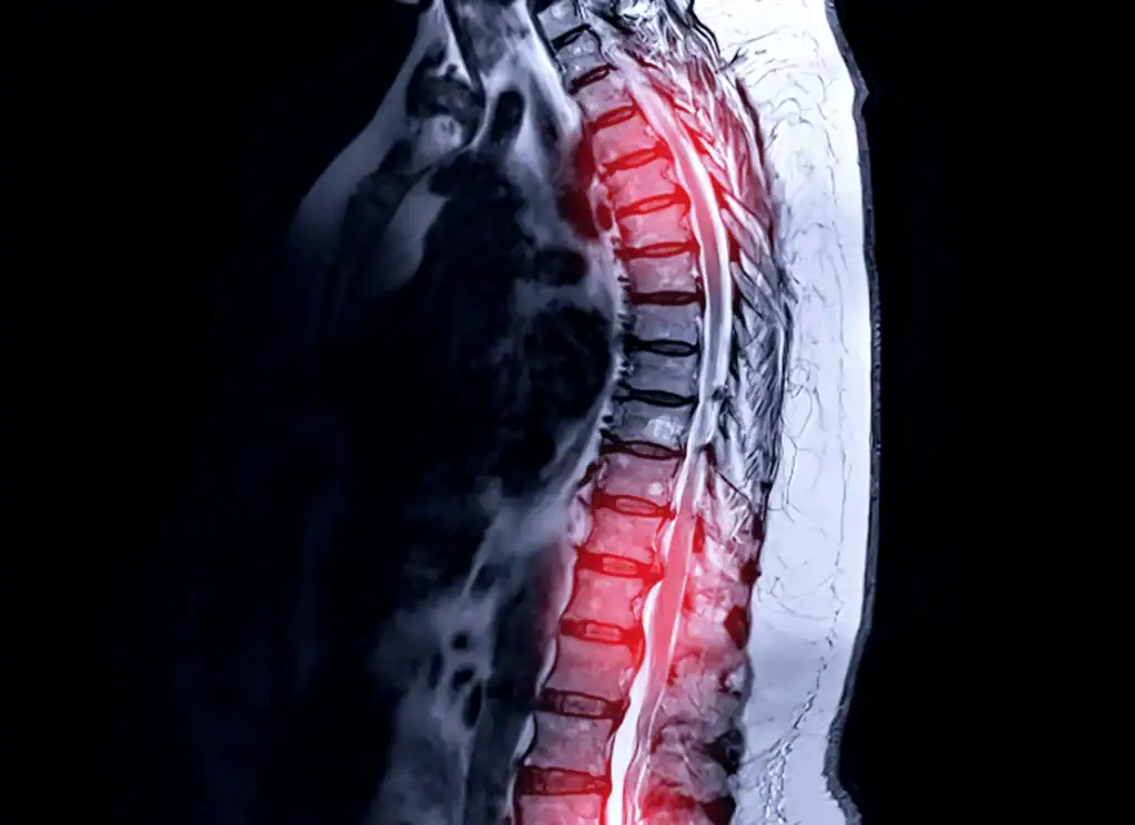
The BioAtlas protocol captures multiple imaging planes throughout the body, including cross-sectional views in key areas such as the spine.

The difference can be significant. A study indexed by the National Center for Biotechnology Information found that sensitivity for spinal canal narrowing was 55% when the spine was viewed from the side alone, compared with 80% when cross-sectional views were also included.

BioAtlas also takes a deeper approach to the spine itself. The protocol includes imaging that evaluates inflammation and fluid, bone marrow, discs and nerve-related structures. Bone marrow evaluation is particularly important for identifying areas where normal marrow may have been replaced by abnormal tissue, including metastatic disease. Many other whole-body MRI protocols simplify the spine imaging by omitting sequences that evaluate bone marrow in detail.

If you are undergoing screening before symptoms appear, these distinctions matter. The ability to find a subtle abnormality depends in part on whether the scan was designed to reveal it.

Advanced MRI Can Reveal Changes You Can’t See

BioAtlas also provides information about aspects of your health that may not be apparent from appearance, weight, or routine health measures.

AI-driven analysis assesses liver fat, iron, and early fibrotic inflammation. Body composition analysis measures visceral fat, subcutaneous and muscular fat, and muscle quantity, quality, and balance. These metrics can reveal metabolic changes and hidden health risk even when you otherwise appear healthy.

A first scan can help establish your baseline. Over time, we can track measures such as liver fat, visceral fat, and muscle quality, helping us see whether meaningful aspects of your biology are changing, and if those changes require interventions.

The Images Are Only the Beginning

Whole-body imaging asks a radiologist to evaluate a large amount of anatomy, including subtle and borderline findings. Differences in interpretation can contribute to false positives and unnecessary follow-up.

Radence uses a small, hand-selected group of experienced radiologists trained on the BioAtlas protocol, creating greater consistency in how scans are interpreted and helping reduce variability from one reader to another.

Then the clinical interpretation begins.

Does a finding fit with your medical or family history? Does another piece of testing make it more significant? Has it changed from previous imaging? Does it require another modality to characterize it? Should it be acted on now or monitored over time?

For you, this is the difference between receiving a finding and understanding what that finding actually means for your health.

One Scan Cannot Answer Every Question

BioAtlas provides a detailed view of the body. Some questions call for another type of imaging.

Your cardiovascular risk, for example, may warrant coronary CT angiography to evaluate the coronary arteries. Lung risk may point to Chest CT. Bone health may require DEXA. Cardiac structure and function may be better evaluated with echocardiography.

BioAtlas therefore sits within a multimodal imaging strategy at Radence. Your biology, risk factors, and findings help determine which imaging can provide the information your clinical team needs.

For you, it means choosing imaging based on the clinical question we are trying to answer.

The same approach extends beyond imaging. Results can be considered alongside other layers of your health data to give the clinical team additional context around what a finding may mean.

The question guides the test. Your biology guides the care.

A Radiology Report Is Not a Care Plan

Suppose BioAtlas identifies a small abnormality.

What happens next depends on what it is, what else we know about you, and how much clinical significance it carries. It may need another type of imaging. It may need to be watched for change. It may warrant specialist expertise or treatment.

This is where Radence’s role extends beyond testing.

Your Radence clinical team determines what deserves further investigation and helps establish the appropriate next steps. When your existing physician or a specialist should be involved, we work alongside them rather than replacing them.

That involvement can be hands-on. A Radence clinician may speak directly with your physician or specialist to explain a finding, share the broader context behind it, and help determine what should happen next. We will join you in appointments with your other physicians when having the Radence clinical perspective in the room can help inform the conversation and treatment plan.

We help carry that clinical context across your care team, so you aren’t left taking a radiology report from one doctor’s office to another and explaining the findings yourself.

Radence stays involved, helping connect the right people, coordinate follow-up, and track what happens over time.

Seeing More Matters When You Know What to Do Next

If you are undergoing advanced imaging to look for disease before symptoms appear, the quality of the scan matters. So does everything surrounding it.

You want confidence that the protocol was designed to capture subtle findings and performed consistently. You want experienced radiologists interpreting the images. You want a clinical team considering those findings in the context of everything else known about your health.

And when something is found, you need answers to a different set of questions:

That is where Radence goes beyond testing.

When MRI raises a question it cannot fully answer, we help determine the right next test. When something deserves attention, we bring in your existing physicians or the appropriate specialists and help coordinate the next steps. When something needs to be followed, we continue looking for meaningful change over time.

Seeing more matters when you know what to do next. And when healthcare gets complicated, Radence stays with you.

See how BioAtlas fits into a more complete picture of your health.

BioAtlas gives us a detailed view of what is happening inside your body. Radence connects that information with the rest of your health to understand what matters, what deserves attention, and what should happen next.

Explore the Radence experience

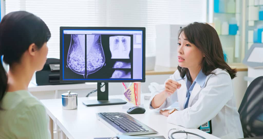
A doctor discuss the results of a mammogram with her patient.

Is a Mammogram Enough?

Breast cancer is the most common cancer among women worldwide. And in the US, a woman has a 1 in 8 chance of being diagnosed with breast cancer in her lifetime. Yet there is hope: When breast cancer is found early, before it spreads, the five-year survival rate is 99%, according to the Centers for Disease Control and Prevention’s US Cancer Statistics on Female Breast Cancer, released in June 2025.

Early screening remains the most powerful tool we have to save lives.

For decades, the mammogram has been the cornerstone of screening. A screening mammogram also serves as an essential first step in determining whether or not a woman has dense breast tissue. This is significant because, for women who have dense breast tissue, a mammogram alone may not be enough.

The Hidden Challenge of Dense Breast Tissue

Dense breast tissue describes the appearance of breast tissue on imaging and occurs when there is a higher concentration of fiber or glands in the breast.

About 4 out of 10 women have dense breast tissue, which is normal, though it can complicate screening. On a mammogram, dense tissue can appear similar to tumors, making abnormalities harder to see. Additionally, studies have shown that women with dense breast tissue have a higher risk of developing breast cancer.

Even a 3D mammogram can miss cancers when dense tissue is present. Dense areas may not cover the whole breast. They can be just in sectors. That means a mammogram could look normal, but a cancer is hiding.

If you have dense breast tissue, an otherwise unremarkable mammogram does not guarantee that you are in the clear.

When Mammograms Aren’t Enough

Magnetic Resonance Imaging (MRI) is technology which uses magnets to generate pictures. MRI does not expose a person to ionizing radiation. For women with dense breast tissue, breast MRI provides another layer of protection, offering a more detailed view. With contrast enhancement, a breast MRI can detect small lesions that mammography may miss.

For women with dense breast tissue, contrast-enhanced MRI can be a game-changer. It can detect cancers at their earliest, most treatable stage.

How to Make Early Detection a Priority

Breast cancer awareness is about knowledge, empowerment, and action. By understanding the role of breast density and family history, women can make more informed choices — and catch cancer earlier, when it’s most treatable.

Here’s what you can do:

Schedule your mammogram. It remains the first step in detection. Ask about your breast density. If your report says you have dense breasts, talk to your doctor about next steps. Know your family history. It is important to know if a close relative was diagnosed with breast cancer or ovarian cancer. In cases of breast cancer, if possible, find out how old your relative was at the time of diagnosis. And ask your doctor if you need to begin screening at an earlier age, even 10 years earlier than the age your relative was when he or she was diagnosed. Advocate for yourself. If you feel you’re at higher risk, don’t hesitate to ask about advanced imaging like breast MRI.

Being proactive about your health with early detection screening may lead to better outcomes. It’s about taking steps that could save your life.

Why It Matters

The difference between finding cancer early and finding it late is profound. When breast cancer is detected locally, before it spreads to other parts of the body, there is a significantly greater chance of survival. Once breast cancer progresses, treatment is more complex, and outcomes become worse.

Dense breast tissue is common, and so is breast cancer. What should be uncommon are missed opportunities for early detection.

Mammograms remain indispensable. But they are not perfect. If you have dense breast tissue — and many women do — a mammogram may not be enough on its own. It is important to know your screening options, and talk with your doctor about the best ways to detect breast cancer.